PET technology expands access and disease detection for oncology and cardiology patients
Nothing can replace the caring touch of a healthcare worker, nor the expert clinical decision-making of a physician or advanced practice provider. But in today’s healthcare environment, technology is equally essential in providing diagnostic analysis and life-preserving care.
At LMH Health, advances in patient-centered technology have transformed the healthcare journey for many patients. Securing leading-edge, innovative equipment has led to more precise diagnosis, greater personalization for surgical implants, and improved treatments and greater healing.
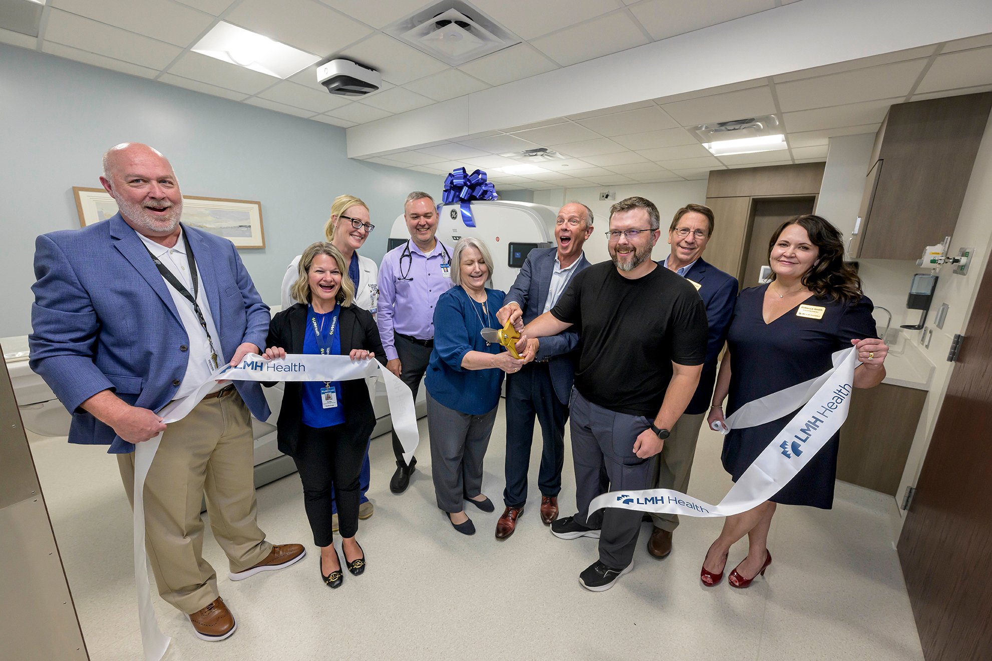
The acquisition of a state-of-the-art, on-campus Positron Emission Tomography (PET) scanner provides these transformative healthcare services to the communities that we serve. Thanks to gifts by donors and corporate partners, including funds raised through the LMH Health Foundation’s biennial Hearts of Gold gala in April and upcoming Penny Jones Golf Tournament, the permanent PET scanner made its debut with public tours and a ribbon cutting on July 18.
How does PET work?
A PET scan reveals how tissues and/or organs are metabolically functioning.

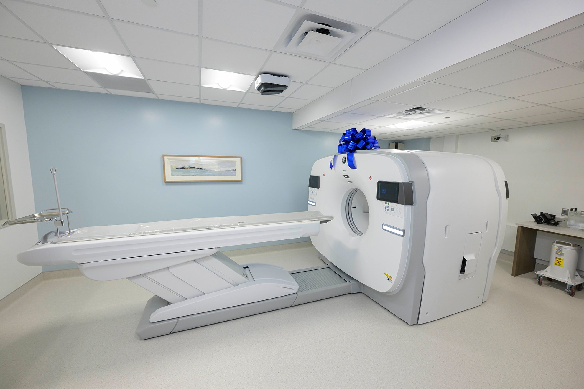
GE Omni PET-CT scanner at LMH Health
“Cancer cells are often metabolically hyperactive,” said Dr. Thomas Grillot, chair of the radiology department at LMH Health and radiologist with Radiologic Professional Services (RPS). “This means they metabolically function differently than healthy, non-cancerous or diseased cells and tissues.”
Patients who need to complete a PET scan are injected with a radioactive medication (also known as a tracer) prior to the procedure. An additional medication can also be given to the patient in advance to help aid in relaxation.
As the patient passes through the large opening of the scanner, the tracer collects in those areas of the body where cells are functioning abnormally, which causes them to appear more prominent on the scanned image to help pinpoint the location of the disease. Indications of diseases typically show up on a PET scan well before other imaging tests such as CT or MRI.
“PET is the gold standard for staging a wide range of cancer diagnoses for treatment; either surgically, through chemotherapy or otherwise,” said Dr. James Mandigo, also a radiologist at LMH Health and with RPS. Dr. Mandigo is fellowship-trained in nuclear radiology, which also happens to be the classification for PET services. “It’s invaluable in evaluating how a tumor is or is not responding to treatment and in determining if the cancer has metastasized to other sites in the body.”
PET and your heart
A PET scan can also be used to evaluate the impact of heart damage after a cardiac event, like a heart attack, or the progression of heart disease. As mentioned before, a PET scan evaluates how tissues metabolically function when a tracer is injected into the body. Since heart tissue is muscle, it is highly metabolic. If it is diseased or damaged, its images will look markedly different from healthy components on a PET scan.
“The level of accuracy and detail in heart images produced by a PET scan are just second to none,” said Dr. Christina Salazar, cardiologist with Cardiovascular Specialists of Lawrence. “Being able to have that level of specificity to determine the amount and extent of damage to heart muscle, areas of reduced blood flow, and whether we need to recommend surgery or other procedures/treatments is invaluable to my patients at risk for heart disease, post heart attack or in determining the progression of a patient’s heart condition.”
Mobile vs permanent
LMH Health utilized a mobile PET scan service for years. Housed inside a large semi-truck trailer, patients walked outside in all kinds of weather to complete their scans in the mobile unit. Availability was limited to one day a week on the Main Campus, and just 16 patients could be scanned in that day.
“We often filled [that schedule] to capacity very quickly,” said Dr. Grillot. “Many weeks, there were more oncology orders for patients than opportunities to scan them in a day. This left little space for non-cancer patients, created delays or inadvertently compelled a patient to travel outside of Lawrence for care.”
The mobile unit is susceptible to weather-related and driver delays, which can postpone imaging until the following week. The travel unit also has a higher operating cost in comparison to a permanently-placed machine, and results could take days to return to the ordering physician.
“Our radiologists are outstanding and read results so timely; sometimes the same day or next day,” said Dr. Sharon Soule, oncologist with the LMH Health Cancer Center. “A permanent PET scanner will expedite evaluation and access to treatment. If we have to send someone to the KC Metro for a PET, results can take several days or up to a week or more.”
Next steps for PET

Ribbon cutting for the PET-CT scanner on July 18, 2024
Construction on the permanent space for the PET scanner began March 25 and was completed in mid-July.
At the outset, PET scans will be available for certain oncology and cardiology patients, with a provider referral. In the future, LMH Health hopes to expand testing to prostate cancer and specific neurological conditions and diseases.
For more information about PET, visit lmh.org/pet. Cost should never be a barrier to care. Financial assistance options are available for persons who are uninsured or underinsured. To learn more, contact LMH Health Patient Accounts at 785-505-5775.